
提供者:上海笃玛生物科技有限公司 发布时间:2024/8/30 14:06:04 阅读次数:2733次 进入该公司店铺
| Product Name | Grp78 antibody , Internal |
|---|---|
| Antibody Type | Primary Antibodies |
| Product description |
|
| Immunogen | Recombinant protein encompassing a sequence within the center region of human Grp78. The exact sequence is proprietary. |
| Clonality | Polyclonal |
|---|---|
| Isotype | IgG |
| Host Species | Rabbit |
| Tested Applications | ICC/IFIHC-PIPWB |
WB:1:100~1:400 More IP:1:20~1:100 ICC/IF:1:20~1:100 IHC-p:1:10~1:50 | |
| Species Reactivity | HumanMouseRat |
| Predicted Reactivity | Zebrafish, Japanese medaka, Xenopus laevis, Chicken, Rhesus Monkey, BovineZebrafish (98%), Japanese medaka (97%), Xenopus laevis (97%), Chicken (100%), Rhesus Monkey (100%), Bovine (100%) |
| Concentration | 0.2 mg/ml |
| Purification | Affinity purified |
| Gene Synonyms | More |
|---|---|
| Target | Grp78 |
| Molecular Weight(MW) | 72 kDa (note) |
| Tissue Specificity | Knockdown/Knockout validation was supported by customer review data. |
| Cellular Localization | Endoplasmic reticulum lumen , Melanosome |
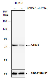
Non-transfected (–) and transfected (+) HepG2 whole cell extracts (30 µg) were separated by 7.5% SDS-PAGE, and the membrane was blotted with Grp78 antibody [N2C1], Internal diluted at 1:200. The HRP-conjugated anti-rabbit IgG antibody was used to detect the primary antibody.
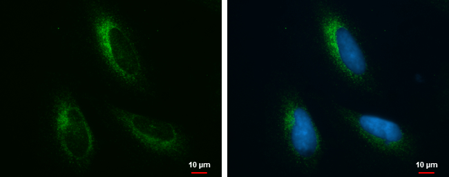
Grp78 antibody [N2C1], Internal detects Grp78 protein at endoplasmic reticulum by immunofluorescent analysis. Sample: HeLa cells were fixed in ice-cold MeOH for 5 min. Green: Grp78 protein stained by Grp78 antibody [N2C1], Internal diluted at 1:100. Blue: Hoechst 33342 staining.
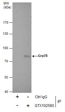
Immunoprecipitation of Grp78 protein from HepG2 whole cell extracts using 5 μg of Grp78 antibody [N2C1], Internal . Western blot analysis was performed using Grp78 antibody [N2C1], Internal . EasyBlot anti-Rabbit IgG was used as a secondary reagent.
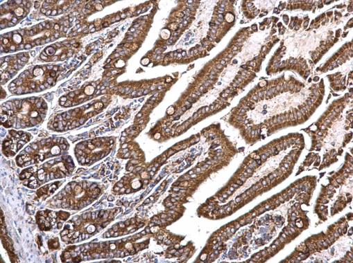
Grp78 antibody [N2C1], Internal detects Grp78 protein at cytosol on mouse intestine by immunohistochemical analysis. Sample: Paraffin-em
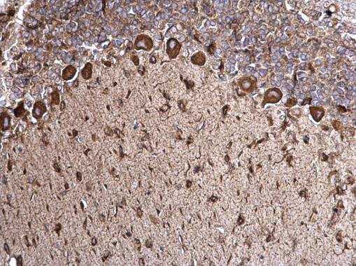
Grp78 antibody [N2C1], Internal detects Grp78 protein at cytosol on rat hind brain by immunohistochemical analysis. Sample: Paraffin-em
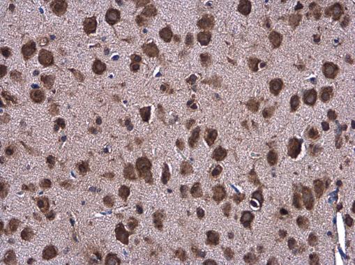
Grp78 antibody [N2C1], Internal detects Grp78 protein at cylasm in mouse brain by immunohistochemical analysis. Sample: Paraffin-em
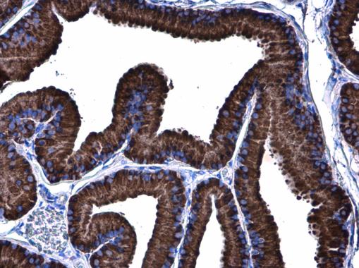
Grp78 antibody [N2C1], Internal detects Grp78 protein at cylasm in rat prostate by immunohistochemical analysis. Sample: Paraffin-em
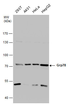
Grp78 antibody detects Grp78 protein by western blot analysis. Various whole cell extracts (30 µg) were separated by 7.5% SDS-PAGE, and the membrane was blotted with Grp78 antibody diluted at a dilution of 1:200. The HRP-conjugated anti-rabbit IgG antibody was used to detect the primary antibody.
| Positive Control | 293T , A431 , HeLa , HepG2 , Neuro 2A , C8D30 , NIH-3T3 , Raw264.7 , C2C12 , PC-12 , Rat2 |
|---|---|
| Application Notes | WB:1:100~1:400 More IP:1:20~1:100 ICC/IF:1:20~1:100 IHC-p:1:10~1:50 |
| Form | Liquid |
|---|---|
| Storage Instructions | Keep as concentrated solution. Aliquot and store at -20ºC or below. Avoid multiple freeze-thaw cycles. |
| Storage Buffer | 1XPBS, 1% BSA, 20% Glycerol (pH7). 0.025% ProClin 300 was added as a preservative. |
① 凡本网注明"来源:阿仪网"的所有作品,版权均属于阿仪网,未经本网授权不得转载、摘编或利 用其它方式使用。已获本网授 权的作品,应在授权范围内使用,并注明"来源:阿仪网"。违者本网将追究相关法律责任。
② 本信息由注册会员:上海笃玛生物科技有限公司发布并且负责版权等法律责任。


版权声明