
提供者:上海笃玛生物科技有限公司 发布时间:2024/8/20 13:54:29 阅读次数:2728次 进入该公司店铺
| Product Name | CD31/PECAM-1 [7-A1] |
|---|---|
| Antibody Type | Primary Antibodies |
| Product description |
|
| Immunogen | Synthetic peptide (KLH-coupled) within C-terminal residues of human CD31. |
| Clonality | Monoclonal |
|---|---|
| Isotype | IgG2a |
| Host Species | Mouse |
| Tested Applications | WBICC/IFIHCFC |
WB:1:1,000-1:5,000 More ICC:1:50-1:200 IHC:1:1,000-1:5,000 FC:1:50-1:100: | |
| Species Reactivity | HumanMouse |
| Concentration | 2 mg/mL. |
| Alternative Names | Hide |
|---|---|
| Molecular Weight(MW) | 82kDa(Observed: 130 kDa) |
| Cellular Localization | Cell junction. Cell membrane. Membrane. |
| SwissProt ID | P16284 Q08481 |
|---|
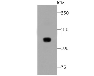
Fig1: Western blot analysis of CD31 on THP-1 cells lysates using anti-CD31 antibody at 1/1,000 dilution.
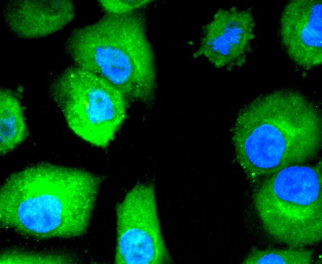
Fig2: ICC staining CD31 in HUVEC cells (green). The nuclear counter stain is DAPI (blue). Cells were fixed in paraformaldehyde, permeabilised with 0.25% Triton X100/PBS.
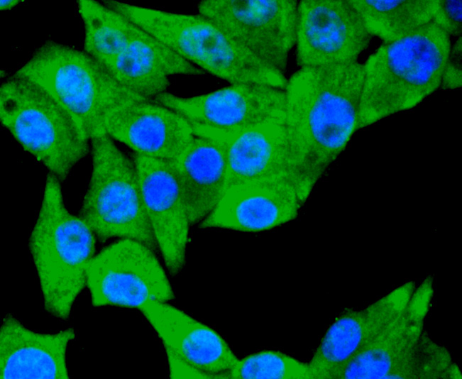
Fig3: ICC staining CD31 in PMVEC cells (green). The nuclear counter stain is DAPI (blue). Cells were fixed in paraformaldehyde, permeabilised with 0.25% Triton X100/PBS.
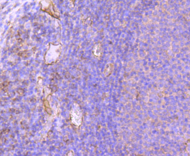
Fig4: Immunohistochemical analysis of paraffin-embedded human tonsil tissue using anti-CD31 antibody. Counter stained with hematoxylin.
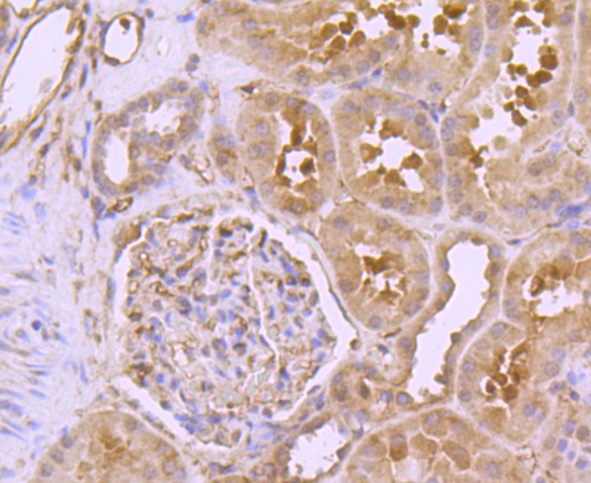
Fig5: Immunohistochemical analysis of paraffin-embedded human kidney tissue using anti-CD31 antibody. Counter stained with hematoxylin.
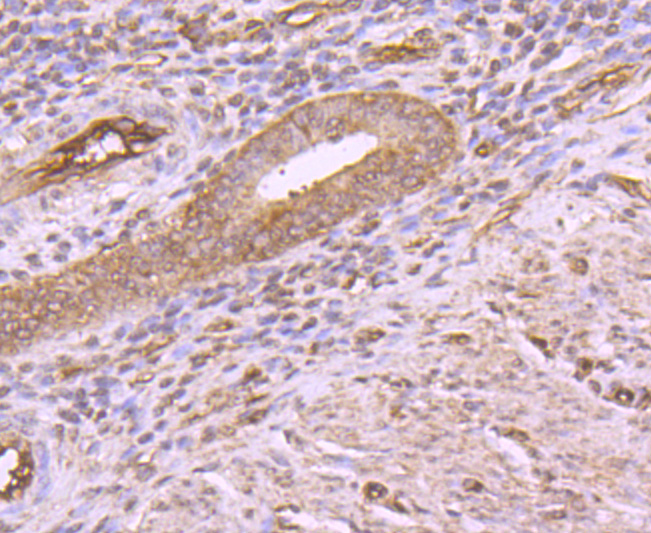
Fig6: Immunohistochemical analysis of paraffin-embedded human uterus tissue using anti-CD31 antibody. Counter stained with hematoxylin.
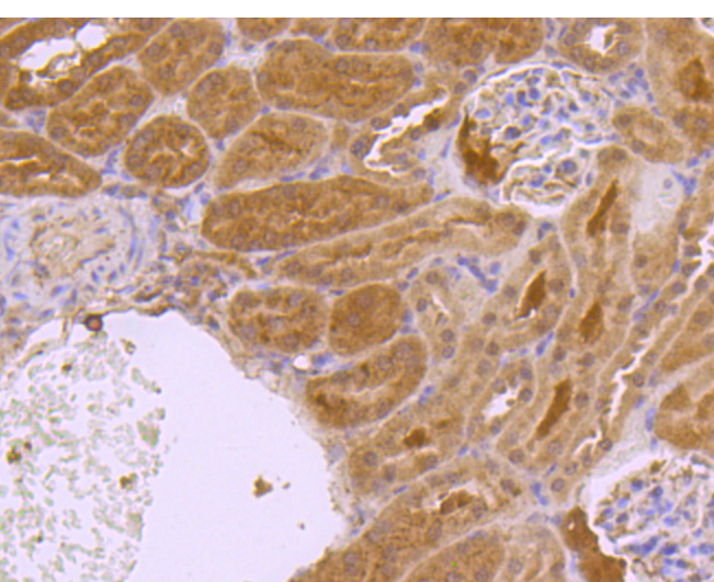
Fig7: Immunohistochemical analysis of paraffin-embedded mouse kidney tissue using anti-CD31 antibody. Counter stained with hematoxylin.
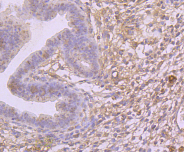
Fig8: Immunohistochemical analysis of paraffin-embedded mouse uterus muscle tissue using anti-CD31 antibody. Counter stained with hematoxylin.
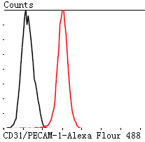
Fig9: Flow cytometric analysis of Jurkat cells with CD31 antibody at 1/100 dilution (red) compared with an unlabelled control (cells without incubation with primary antibody; black). Alexa Fluor 488-conjugated Goat anti mouse IgG was used as the secondary antibody.
| Positive Control | THP-1, HUVEC, PMVEC, NIH/3T3, Jurkat, MCF-7, human tonsil tissue, human kidney tissue, human uterus tissue, mouse kidney tissue, mouse uterus tissue |
|---|---|
| Application Notes | WB:1:1,000-1:5,000 Hide ICC:1:50-1:200 IHC:1:1,000-1:5,000 FC:1:50-1:100: |
| Form | Liquid |
|---|---|
| Storage Instructions | Store at +4℃ after thawing. Aliquot store at -20℃. Avoid repeated freeze / thaw cycles. |
| Storage Buffer | 1*TBS (pH7.4), 1%BSA, 40%Glycerol. Preservative: 0.05% Sodium Azide. |
① 凡本网注明"来源:阿仪网"的所有作品,版权均属于阿仪网,未经本网授权不得转载、摘编或利 用其它方式使用。已获本网授 权的作品,应在授权范围内使用,并注明"来源:阿仪网"。违者本网将追究相关法律责任。
② 本信息由注册会员:上海笃玛生物科技有限公司发布并且负责版权等法律责任。


版权声明