上海笃玛生物科技有限公司
 第12年
第12年


当前位置:阿仪网 > 上海笃玛生物科技有限公司 > 技术文献 > CD31 antibody Catalog Number:OM244946
发布时间:2024/8/20 13:46:25关注度:2711
| Product Name | CD31 antibody |
|---|---|
| Antibody Type | Primary Antibodies |
| Product description |
|
| Immunogen | Human PECAM-1 |
| Clonality | Monoclonal |
|---|---|
| Isotype | IgG2a |
| Clone Number | HEC7 |
| Host Species | Mouse |
| Tested Applications | ELISAFACSGSAICC/IFIHCIPWB |
| WB:1:500~1:2000 Hide IP:1:100~1:500 ICC/IF:1:50~1:100 IHC:1:50~1:200 | |
| Species Reactivity | BovineHumanRat |
| Concentration | 1 mg/ml (Please refer to the vial label for the specific concentration) |
| Purification | Protein G |
| Gene Synonyms | Hide |
|---|---|
| Target | CD31 |
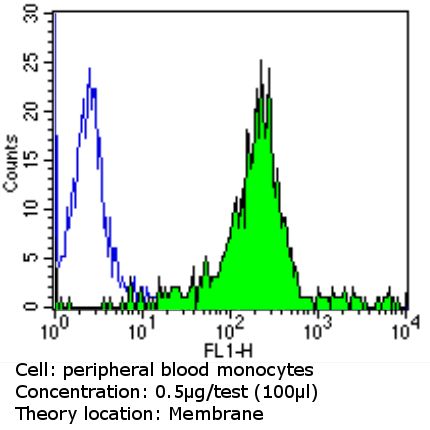
FACS analysis of PBMC cells using CD31 antibody [HEC7] (green) or an isotype control (blue).

FACS analysis of PBMC cells using CD31 antibody [HEC7].
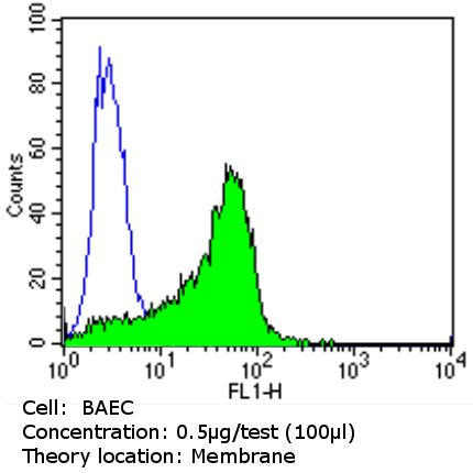
Flow cytometry analysis of CD31 in BAEC cells (green) compared to an isotype control (blue). Cells were harvested, adjusted to a concentration of 1-5x10^6 cells/ml, fixed with 2% paraformaldehyde and washed with PBS. Cells were blocked with a 2% solution of BSA-PBS for 30 min at room temperature and incubated with CD31/PECAM-1 antibody [HEC7] at a dilution of 0.5 ug/test for 60 min at room temperature. Cells were then incubated in a proper secondary antibody and re-suspended in PBS for FACS analysis.
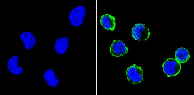
Immunofluorescent analysis of CD31 (green) in THP-1 cells (right) compared to a negative control without primary antibody (left). Formalin-fixed cells were permeabilized with 0.1% Triton X-100 in TBS for 5-10 minutes and blocked with 3% BSA-PBS for 30 minutes at room temperature. Cells were probed with CD31/PECAM-1 antibody [HEC7] in 3% BSA-PBS at a dilution of 1:100 and incubated overnight at 4°C in a humidified chamber. Cells were washed with PBST and incubated with a proper secondary antibody in PBS at room temperature in the dark. F-actin (red) was stained with a flourescent red phalloidin and nuclei (blue) were stained with Hoechst or DAPI. Images were taken at a magnification of 60x.
| Application Notes | WB:1:500~1:2000 Hide IP:1:100~1:500 ICC/IF:1:50~1:100 IHC:1:50~1:200 |
|---|
| Form | Liquid |
|---|---|
| Storage Instructions | -20°C |
| Storage Buffer | PBS |
原创作者:上海笃玛生物科技有限公司
相关信息
推荐产品
ELISA试剂盒,动物血清,生化试剂盒,标准品,对照品,试剂,耗材,抗体,培养基,代理进口品牌,实验代测服务

 在线咨询5分钟应答